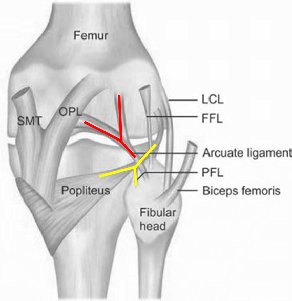
Posterolateral corner (PLC)
It is a complex unit responsible for posterolateral knee joint stabilization. The unit consists of:
- Biceps femoris tendon (BT)
- Popliteus tendon (PT)
- Lateral collateral ligament (LCL) = fibular collateral ligament (FCL)
- Popliteofibular ligament (PFL)
- Posterolateral capsule
- Arcuate ligament (AL)
- Fabellofibular ligament (FFL)
In other words, they are all of the structures in the lateral collateral complex, excluding the iliotibial (IT) band, and can be injured during PLC injuries.
PLC Injuries
Mechanism of injuries
- Hyperextension (Most common)
- Contact
- Non-contact
- Pivot shift: External rotation direct trauma to the anteromedial aspect of the knee
- Noncontact varus force to the knee (varus overload)
- Direct posterior tibial translation (dashboard injury)
Associated injuries
Injuries to the PLC rarely occurs in isolation. Because of the underlying mechanism of injuries, it is typically associated with
- Cruciate ligaments
- Both menisci
- Medial collateral ligament (MCL)
PLC instability can lead to failure of ACL reconstruction and acceleration of patellofemoral OA. Severe PLC injury can also present with foot drop due to peroneal nerve injury because of the nerve’s proximity to the fibular head and LCL insertion.
Investigation
MRI is investigation of choice in patients clinically suspected of having posterolateral corner injuries.
Evaluation
PFL and BT
PFL is a small but important ligament that normally extends from the popliteus tendon and conjoins with the biceps femoris tendon at the insertion on the lateral aspect of the head of the fibula. It is significant for the stability of the PLC and is best seen on coronal and sagittal images.
In sagittal view, PFL can be seen as comma-shaped structure as it extend from the popliteus tendon.
Arcuate sign
Avulsion fracture of the lateral aspect of the head of the fibula at the insertion of the conjoined tendon of the FCL and BT.
FCL
Arising from the lateral surface of lateral femoral condyle and attaching distally onto the fibular head.
PT
Arising from the lateral surface of lateral femoral condyle from a sulcus just inferior to the FCL origin and its muscle belly extending inferomedially beneath the gastrocnemius muscles.
Injury to the muscle or proximal myotendinous junction (MTJ) is very common. So, isolated popliteus muscle injury is not associated with posterolateral instability.
AL
It is a triangular fascial plane draping over the PT and PFL. It separates from the oblique popliteal ligament (OPL) and inserts at the medial aspect of the head of the fibula. It has two bundles, medial and lateral, which are not easily visualized on MRI.
Fabella and FFL
The fabella is an ovoid ossicle that articulates with the posterior lateral femoral condyle. It is a sesamoid of the gastrocnemius, presenting in about 25% of knees. Its importance is still controversial. The FFL is a thin ligament that extends from the ossicle to the fibula head.

Leave a Reply