Overview
One common cause of arthritis. Cause by intra-articular inflammation induced by crystal deposits.
Most common crystal deposits are
- Calcium pyrophosphate → CPPD
- Monosodium urate → Gout
- Calcium hydroxyapatite → Calcific tendinitis
Calcium pyrophosphate (CPP)
- Causing CPPD arthritis (Pseudogout)
- Positively birefringent rhomboid crystal
- Radiographic hallmark finding = Chondrocalcinosis. On radiography and CT, chondrocalcinosis of the articular cartilage can be seen as thin, linear calcifications parallel to the subchondral bone plate. However, this finding may not be the cause of patient pain nor be seen in early CPPD.
- Acute CPP arthritis may mimic septic arthritis, Chronic CPP can mimic almost any arthropathies
- Can deposit in ligaments and synovium, appears as an indistinct, cloud-like mineralization.
Notable manifestations
- Wrist: SLAC can be caused by severe TFCC chondrocalcinosis due to destruction of the scapholunate ligament.
- Knee: CPP arthritis of the knee usually affects the patellofemoral compartment first before progress to involve all three compartments. DDx: patellar maltracking.
- Hand: typically involved radial aspect of carpal head of the 2nd and 3rd MCP joints. DDx: hemochromatosis.
Monosodium urate (MSU)
- Causing gouty arthritis
- Negatively birefringent needle-like crystals within neutrophils due to excess uric acid
- Undersecretion (90%): mostly due to renal insufficiency
- Overproduction (10%)
- Radiographic hallmark finding = sharply marginated erosions with overhanging edges.
- Joint spaces are typically preserved until late stage.
- Ultrasound findings = double contour sign which represent an irregular hyperechoic line of MSU deposited on the hyperechoic cartilage.
- Tophi = Nodules consist of MSU + giant cells + inflammatory cells in soft tissues. Likely to occur in the olecranon bursa of the elbow but can develop anywhere, including within the joint.
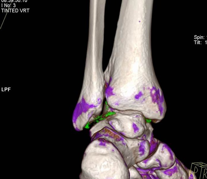
- Dual-energy CT can be used to identify MSU crystals. Post-processing color code is typically green.
Calcium hydroxyapatite
- Most common subset of basic calcium phosphate (BCP) crystals.
- Causing calcific tendinitis. More commonly seen in DM patients.
- Radiograph findings = amorphous or globular calcification. Absent of cortication or internal trabeculation helps distinguish it from conditions that have bone formation by osteoblasts eg., intra-articular body, fragmented enthesophyte, or osteochondromatosis.

Leave a Reply