Introduction
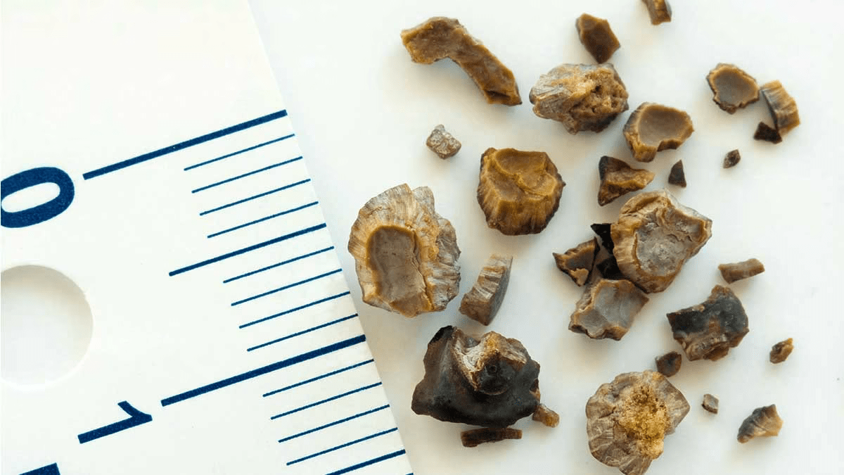
Did you know that 1 in 11 people in the U.S. suffer from the excruciating pain of urolithiasis, or kidney stones? This condition not only causes immense discomfort to those affected but also places a significant burden on our emergency departments, with nearly a million visits each year. While many cases resolve themselves through the spontaneous passage of the stones, others present as emergencies requiring procedural interventions. Let’s delve into what makes kidney stones such a daunting challenge for both individuals and healthcare systems.
Classifications
Urolithiasis is categorized based on its location within the urinary tract and its chemical composition, with each aspect significantly influencing management strategies. The etiology varies across different types of stone compositions, leading to distinct preventive measures for each.
By location
- Upper urinary tract
- Calyceal
- Renal pelvis
- Ureteropelvic junction (UPJ)
- Ureteral calculi
- Ureter
- Ureterovesicle junction (UVJ)
- Lower urinary tract
- Urinary bladder
- Urethral
By composition
From the most common to the less common in descending order.
- Calcium (75-80%)
The most common type of urolithiasis by composition is predominantly made up of calcium oxalate, although some calcium stones are composed of calcium phosphate. - Struvite (15-20%)
Magnesium ammonium phosphate ± calcium phosphate (triple phosphate) - Uric acid (5-10%)
- Cystine (1-3%)
- Matrix (rare): mucoproteins
- Xanthine (extremely rare)
- Protease inhibitor (Indanivir induced)
- Milk of calcium: carbonate apatite
Most stones are mixed composition with more than half contain calcium salts.
Epidemiology
The incidence of urolithiasis peaks in young adults aged 20-40 years, with a rate of about 1-2 per 1000 individuals.
The most common type, calcium stones, are found more frequently in men, with a male-to-female ratio of 3:1.
Conversely, struvite and matrix stones are more commonly seen in women, with a male-to-female ratio of 1:2.5.
Etiology
Composition
Etiology
Calcium
(Radiopaque)
- Idiopathic (85%): Idiopathic hypercalciuria
- Acquired (15%)
- Hyperparathyroidism
- Sarcoidosis
- Renal tubular acidosis
- Hyperoxaluria
- Steroids
- Cushing syndrome
- Immobilization
- ↑ Vitamin D
Struvite
(Radiopaque)
- Urinary tract infection
- Proteus milabilis
- Klebsiella pneumonaie
- Escherichia coli
- Pseudomonas aeruginosa
Uric acid
(Radiolucent)
- Hyperuricosuria
- 25% with Gout
- Ileostomy
- Chemotherapy
- Acidic and concentrated urine
- Adenine phosphoribosyltransferase deficiency
Cystine
(Radiolucent)
- Cystinuria (autosomal recessive; SLC3A1, SLC7A9)
Matrix
(Radiolucent)
- Chronic UTI
- Urine obstruction/stasis
Milk of calcium
(Radiopaque)
- Calyceal diverticula
- Ureteroceles
Risk factors
Intrinsic
- Prior history of urolithiasis
- Personal
- Family
- Anatomical abnormalities
- UPJ obstruction
- Horseshoe kidney
- Ectopic kidney
- Pelvocaliceal diverticula
- Tubular ectasia
- Urinary diversion
- UPJ obstruction
- Malabsorption (↑ urinary oxalate)
- Roux-en-Y gastric bypass
- Sleeve gastrectomy
- Medical conditions(4)
- Chronic kidney disease
- Hypertension
- Gout
- Diabetes mellitus
- Hyperlipidemia
- Obesity
- Endocrine disorders
- Malignancy
Extrinsic
- Lifestyle
- Environment
- warm climates
- summer
- Medications
- Acetazolamide
- Sulfadiazine
- Ceftriaxone (prolonged used)
- Protease inhibitors (HIV medication)
- Indinavir
- Atazanavir
In the prevention section, we will discuss strategies involving the mitigation of risk factors.
Pathogenesis
Stones formation
There are two theories that describe the growth and aggregation of crystal.
- Fix particle mechanism (Anderson-Carr-Randall theory)
- Free particle mechanism
1. Fix particle mechanism
Composition of crystals supersaturation in the renal medulla
⬇︎
Microaggregates increase in size
⬇︎
Migration toward caliceal epithelium
⬇︎
Rupture into calices to form calculi
The fixed particle mechanism states that stones formed attached to calcific plaques called Randall plaques. These plaques can be visualized as a thin (<2 mm) calcified plaques at renal papillae on CT imaging.
2. Free particle mechanism
Composition of crystals supersaturation in the urine within the tubules
⬇︎
Microaggregates increase in size within tubules
⬇︎
Obstruction of urine outflow
⬇︎
Formation of more stones
Across both fixed and free mechanisms, the formation of microaggregate crystals is proposed to occur through three distinct processes.
- Nucleation
Crystal/foreign body initiates formation in urine supersaturated with crystallizing salt - Stone matrix
Organic matrix of urinary proteins + serum serves as framework for deposition of crystals - Inhibitor
Little/no concentration of urinary stone inhibitors such as- Citrate
- Pyrophosphate
- Glycosaminoglycan
- Nephrocalcin
- Tamm-Horsfall protein
Mechanism of renal colic
Stones initially form in the kidneys
(Nephrolithiasis)
⬇︎
Distally migrate into the ureter
(Ureteric calculi)
⬇︎
Obstruction of urine outflow
⬇︎
Upstream dilatation of the ureter (hydroureter) and renal pelvocaliceal system (hydronephrosis)
⬇︎
Increase luminal tension
⬇︎
Prostaglandin release
⬇︎
Ureteral peristalsis and colicky pain
Clinical manifestation
Regardless of the stone type, patients may exhibit a range of symptoms, from being asymptomatic to critically ill.
Factors that affect clinical findings include the site/degree of obstruction and stone size.
Site of urolithiasis
Upper UT
- Asymptomatic
- Flank pain
- Fever ± sepsis
Even small, non-obstructing, renal stones can be symptomatic due to intermittent obstruction or epithelial irritation.
Ureter
Acute colicky pain radiating to the groin
Common sites of obstruction (More to less)
- Ureteropelvic junction (UPJ)
- Distal ureter (at iliac crossing)
- Ureterovesical junction (UVJ)
Lower UT
- Asymptomatic
- Dysuria
- Dull/sharp pain radiating to genital/buttocks/perineum
UVJ stones may cause similar clinical findings as lower UT stones.
Associated symptoms
Due to prostaglandin release, there may be associated symptoms such as;
- nausea
- vomiting
- fever
Complication
In cases of severe and/or prolonged urinary obstruction. There may be;
- (Post-renal) acute renal failure
- Oliguria/anuria
- Urinary tract infection
- Sepsis
Laboratory findings
- microscopic hematuria in urinalysis (may be absent in acute total obstruction)
- ↑ serum Cr (due to obstructive uropathy)
Choice of imaging modality
Ultrasound KUB
Pros
- No radiation
→ imaging study of choice in pediatric and pregnant patients - Can identify hydronephrosis secondary to obstructive urolithiasis
- Can assess urinary flow via doppler jet.
Cons
- Limit evaluation of ureteric stones.
(Sn 57%, Sp 97.5%) - Limited in patients with large body habitus
- Operator dependent
Plain film KUB
Pros
- Helpful in monitoring stone growth over time
Cons
- Limit evaluation of radiolucent stones
CT for urinary stones
Pros
- Best accuracy
(Sn 95%, Sp 98%) - May predict lithotripsy response
(↑HU → ↓success)
Cons
- Not visualized
- Protease inhibitor induced stones (Indinavir)
- Matrix stones
- Radiation dose especially in obese patients
MRI for urinary stones
Pros
- No radiation dose → second-line imaging option for pregnant and pediatric patients
- Better accuracy than ultrasound (but inferior to CT)
Cons
- Technical problems
- More expensive
- Time consuming
- Not readily available
Acute management
Conservative medical therapy
Approximately 86% of stones pass spontaneously within 30-40 days, with success rates dependent on size. Therefore, patients with smaller (< 5-6 mm), uncomplicated stones may be managed conservatively.
Pain control
- NSAIDs
- Directly counteract prostaglandin release → first-line treatment for pain
- Can be given as IV or oral
- Opioids
- Reserved for refractory pain
Nausea and vomiting management
IV antiemetic medications such as
- Ondansetron
- Metroclopramide
- Promethazine
Dehydration management
Considered IV crystalloid in patients with sign of moderate to severe dehydration or persistent vomiting.
Caution: Use of IV hydration does not show to facilitate stone passage.
Medical expulsive therapy (MET)
- Alpha-blockers
Useful adjunct to facilitate passage of 5-10 mm stones but not beneficial for smaller ones.- Doxazosin
- Tamsulosin
Hospitalization
Hospitalization for close observation may be considered in patients with;
- Intractable pain or vomiting
- Inability to tolerate oral intake
- Pregnancy
- Pediatric age
Urological intervention
Considered urgent/emergent urological intervention in patients with;
- Larger stones (which are unlikely to spontaneously stone passage)
- Acute complication
- Solitary kidney
Intervention methods
Actual intervention plan should be discussed with urologist according to patient’s presentation, medical conditions, and urologist’s comfort and preference.
Flexible ureteroscopy (URS)
- The most common method used
- Direct endoscopic visualization of the urinary tract and retrieval of an obstructing stone
- Good option for lower pole stones between 1.5-2 cm
- Ideal choice of treatment for patients taking anticoagulant/antiplatelet medication
Extracorporeal shockwave lithotripsy (ESWL)
- Used of ultrasound shockwaves from the body surface to fragment the stone into smaller pieces that can be passed into urine
- May required follow up flexible URS for ureteral stent
- Typically need IV sedation or general anesthesia (but can be performed on an outpatient basis)
- Some stones are resistant to this method
- Cystine stones
- Matrix stones
- ↑HU calcium stones
Percutaneous nephrolithotomy (PCNL)
Preferred in
- Size > 20 mm
- Staghorn calculi
- Kidney and proximal ureter stones
- Chronic kidney disease
Contraindications
- Pregnancy
- Bleeding disorders
- Active urinary tract infections
Emergent decompression
Considered in patients with acute obstruction with signs of urinary tract infection to prevent permanent renal damage and worsening of infection.
- Indwelling ureteral catheter
- Nephrostomy tube
Prevention
Lifestyle modification
Oral hydration
Oral hydration is recommended at a rate that produces approximately 2.5-3.5 L of urine per day.
Acceptable choice of fluid included
- Water
- Coffee
- Tea
- Beer
- Low sugar fruit juices
except for- Tomato (↑Na content)
- Grapefruit (↑Oxalate content)
- Cranberry (↑Oxalate content)
Diet modification
Avoid intake of;
- High oxalate foods(1)
- Fish oil
- Vitamin C
Increase intake of;
Specific stones prevention
Calcium stones
- Medications
- Thiazide diuretics
- Citrate salts (K citrate)
- Lifestyle modifications
Uric acid stones
- Medications
- Citrate salts (K citrate)
- Uric acid lowering medication
- Allopurinol
- Lifestyle modifications
Cystine stones
- Medications
- Citrate salts (K citrate)
- Thiol drugs
- Lifestyle modifications
Differential diagnosis
KUB
- Pyelonephritis
- Renal abscess
- Renal artery aneurysm
- Lower urinary tract infection
GI
- Appendicitis
- Diverticulitis
- Mesenteric ischemia
- Pancreatitis
- Cholesystitis
- Small bowel obstruction
- Constipation
Gyne
- Ovarian torsion
- Dysmenorrhea
- Ectopic pregnancy
- Spontaneous abortion
- Pelvic inflammatory disease
Footnotes
(1) List of high oxalate foods
- Beans
- Beer
- Berries
- Coffee
- Chocolate
- Some nuts
- Some teas
- Soda
- Spinach
- Potatoes
(2) List of high citrate foods
- Lemon
- Orange
- Melon
(3) List of high calcium foods
- Milk
- Tofu
- Orange
- Almonds
(4) Medical conditions
May increase risk of stone formation due to
- Association with diet high animal-derived proteins, salt, and sugar in patients with obesity, hyperlipidemia, hypertension, and type 2 diabetes mellitus
- Insulin resistance in obesity and type 2 diabetes mellitus
- ↑ urinary calcium
- ↑ urinary pH

Leave a Reply