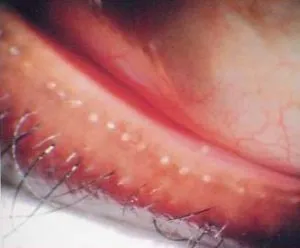
Anatomy and functions
The meibomian gland is a type of sebaceous gland with tubulo-acinar structure and holocrine function, located in the superior and inferior tarsal plates (both posterior eyelids). It secretes lipid compound called “meibum” to ocular surface.
Meibum
Compositions
- Polar lipids
- phospholipids
- Nonpolar lipids
- cholesterol
- wax esters
- cholesterol esters
Functions
- Tear film stability
- Innate microbial resistance
Meibomian gland dysfunction (MGD)
MGD is a type of posterior blepharitis that involves inflammation posterior to the gray line of the lid margin. It is the leading cause of evaporative dry eye and one of the most common conditions encountered by eye care providers. Symptoms of MGD significantly impact quality of life.
Classifications
By rate of gland secretion
- Low delivery state
- hyposecretion
- obstruction (most common)
- cicatricial
- non-cicatricial
- high delivery states
By etiology
- Primary
- Secondary
One diagnosis can be classified in conjunction together. For example;
- Mucus membrane phephigoid = secondary cause of obstructive, cicatricial MGD
- Seborrheic dermatitis = secondary cause of both obstructive non-cicatricial and hypersecretory MGD.
Pathophysiology
Since low deliver states due to gland obstruction is the most common cause of MGD in clinical practice. We will only focus its pathophysiology in this topic which includes:
- Epithelial hyperkeratinization
- Meibum stasis
- Cystic dilation
- Eventual disuse acinar atrophy
- Gland dropout
Newer studies describe meibocyte abnormalities as a contributing mechanism in MGD. MGD risk factors effecting meibocytes differentiation and renewal includes:
- Intrinsic factor (Aging)
Decrease expression of PPAR? (peroxisome proliferator-activated receptor gamma) - Extrinsic factor (Environmental stress)
Risk factors of MGD
Hormonal
Estrogens: increase inflammation
Androgens: stimulate meibum secretion + suppress inflammation
Conditions with androgen deficiency associated with MGD include:
- Anti-androgen medications: (for BPH and CA prostate)
- complete androgen insensitivity syndrome
- Sjogren’s syndrome
Systemic medications
- 13-cis-retinoid acid (Accutane)
Severe atrophy of the meibomian glands. - Carbonic anhydrase inhibitors
Topical medication
- Topical epinephrine
- Topical beta blocker
- Topical prostaglandin analogs
Dietary intake
Supplementation with omegas decreases ocular surface inflammation in patients with dry eye, probably due to alterations in polar lipid profile and decreases in the saturated fatty acid content of meibum.
Ocular surface microbiome
Cholesterol esters in meibum may stimulate proliferation of commensal organisms, such as Staphylococcus aureus, on the eyelid margin.
Commensal organisms → lipolytic enzymes → breakdown of fats and releasing of glycerides and fatty acids (polar lipid) into tear film → diffusion of polar lipids across aqueous into mucin layer → unstable tear film
Also hyperkeratinization may be associated with lipolytic decomposition.
In recent literature, Demodex mite contributions to MGD is not clear.
Contact lens wear
Probably related with mechanical trauma, plugging due to aggregation of desquamated epithelial cells at gland orifices, and/or chronic inflammation.
Congenital diseases
Meibomian glands can be congenitally decreased or absence in conditions such as:
- Turner syndrome
- Ectrodactyly with ectodermal dysplasia and cleft-lip and palate (ECC syndrome)
- Anhidronic ectodermal dysplastic syndrome
Meibomian glands can be congenitally replaced in conditions such as:
- Dystichiasis (by eyelashes)
- Secondary due to metaplastic reaction in mucocutaneous disease
- Autosomal dominant disorder lymphoedema
Clinical presentations
As per slit lamp examination:
- Meibomian orifice plugging
- Eyelid margin foaminess
- Hyperemia/telangiectasia
- Changes in orifice position with respect of the mucocutaneous junction
- Opaque, viscous and difficult to express meibum
- Meibomian gland dropout (more accurate with infrared photography)
In most patients, signs of meibomian gland dysfunction are not correlated with clinical dry eye. Almost twice the number of people with MGD are asymptomatic compared to symptomatic individuals.
Treatment
The goal of treatment for MGD is to improve the flow of meibomian gland secretions and ultimately help reestablish tear film stability.
There is no gold standard treatment, but a variety of options are available, with warm compression and lid hygiene remaining the first-line treatment.
Medical treatment
Antibiotics
Their uses show reduce growth of lid bacteria which may suppress bacterial lipases and inflammation.
Tetracyclines:
- Doxycycline and minocycline are more lipophilic and more concentrated in ocular and lid tissues at lower doses compare to other tetracyclines.
- Side effects: photosensitivity, GI symptoms
- Contraindicated in pregnant women and children (< 8 years old)
Azithromycin:
- Topical
- Systemic
- Beware of cardiac arrhythmia, notably QT prolongation.
Anti-inflammatory agents
- Cyclosporine A: calcineurin inhibitor
- Lifitegrast: lymphocyte function-associated antigen-1 antagonist
- Corticosteroid: only for acute bouts of inflammatory complications of MGD, such as chalazion, marginal keratitis.
- Anakinra: IL-1 receptor antagonist
Essential fatty acids
Demodex treatment
Procedural treatment
- Expression of meibum
- Intraductal meibomian gland probing
- Electronic heating devices
- Intense pulsed light therapy (IPL)
- Intranasal tear neurostimulation (ITN)
Reference
- Meibomian Gland Disease: The Role of Gland Dysfunction in Dry Eye Disease
- Management of meibomian gland dysfunction: a review
Related authority organization:

Leave a Reply